We’re looked at how augmented reality might possibly affect the field of cardiology right here and now thanks to the INSIGHT HEART, an amazing software we’ve reviewed before from the guys at ANIMA RES. Initially, we said that the INSIGHT HEART was focused heavily on the educational side, and we were a bit bothered by that, knowing that it had the potential to be so much more. However, after we talked with them, we were shown that the INSIGHT HEART’s full potential is really being utilized. You can read more about our interview with them here, and we suggest that you do, because in this article, we’re going to discuss the INSIGHT HEART’s potential for AR in interventional cardiology, specifically for addressing congenital and structural heart disease.

Congenital heart disease (CHD) are relatively common, affecting about 1% of births in the US, or about 40,000 children, according to the CDC. These heart diseases are usually structural malformations, and can be either interauricular or interventricular problems. The most common of these CHDs is what’s called a ventricular septal defect (VSD), and of all children born with VSD, 25% of them will need surgery to correct it within the first year of life because of the severity. If you’re interested in reading more about heart conditions in infants, you can read more about it here.
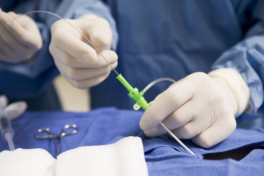
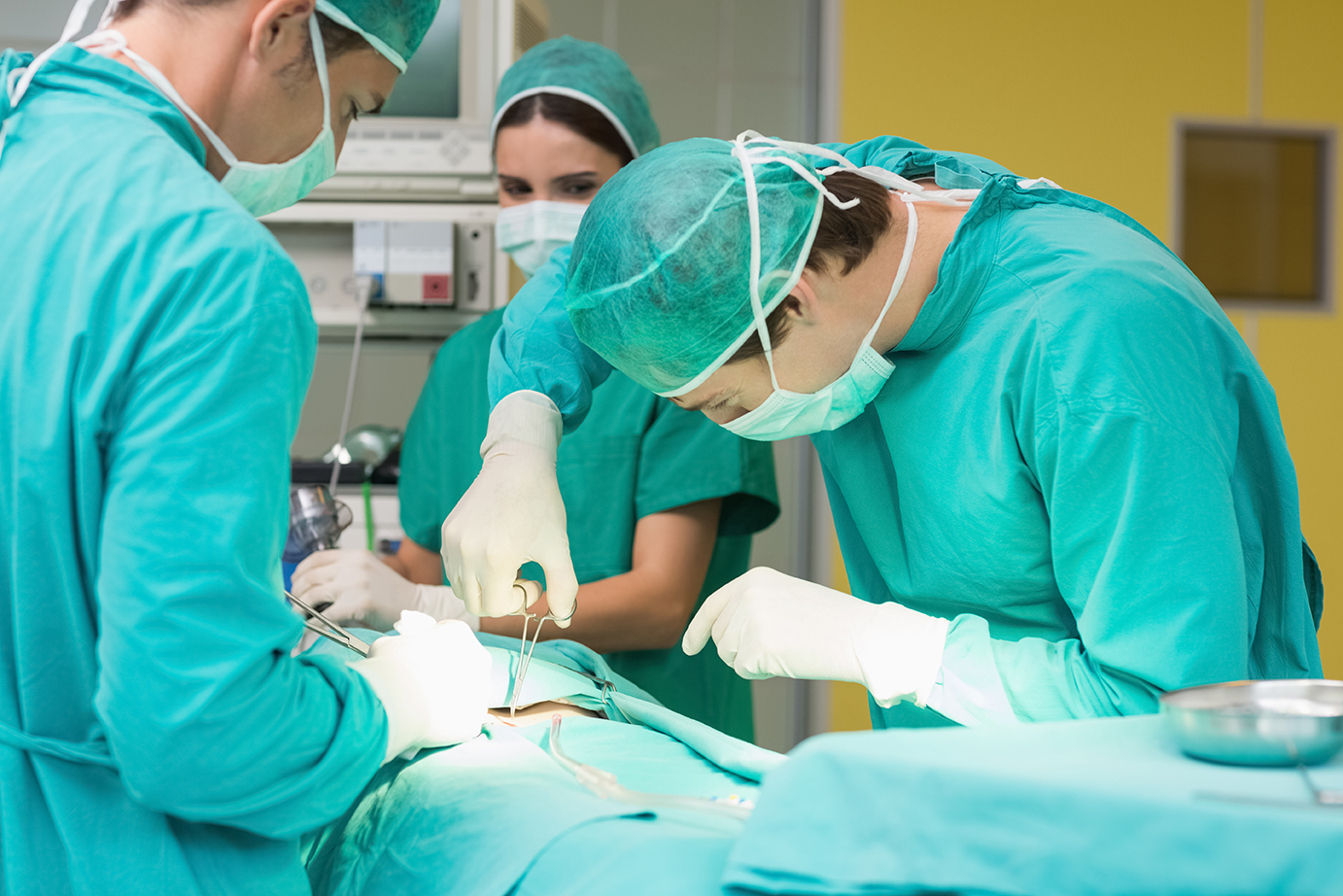
However, when a child’s life is at stake, numbers don’t mean much. What’s really important is to find ways to fight CHD and make life better for children born with it. With this in mind, researchers from the Children’s National Medical Center’s Division of Cardiology in Washington D.C. have explored the potential of AR applications to solving this issue, taking into account that more and more heart defects that previously required open heart surgery can now be addressed in the cardiac catheterization laboratory. As you may imagine: it’s not easy. Cardiac catheterization, especially in newborns, requires a lot of preparation and planning in order to be successful. The images required for the procedure is collected through advanced cardiac imaging such as cardiac magnetic resonance and cardiac computed tomography. The question then becomes “how can we improve upon this technology?”

Augmented Reality looks to answer that question. 3D printed models, though admittedly more expensive, allow the surgeon to prepare for this kind of delicate procedure. It assists cardiologists in a preoperative and operative setting, ranging from using this model to explain the procedure to the parents and answer any questions they might have, since parents are very commonly (and very rightfully) nervous and anxious about allowing their child to undergo surgery. This 3D model can also allow the surgeons to consult each other about the best course of action in a more complex case through a connection through the Microsoft Hololens. They can then discuss each case and decide the best technique before the surgery, or even in an operative setting with the 3D model side by side with the fluoroscopy image, and they can attempt to correct any misjudgements or doubts by sharing notes posted from the preoperative setting in the device used during the surgery.
As with all the other amazing AR tech we talk about here, it sounds very sci-fi, but it’s within our grasp. The future is within our reach, and alternative technologies like AR are saving lives and making medical conditions more bearable for patients everywhere, no matter how young. We hope to hear more from ANIMA RES and their technologies in the future!


