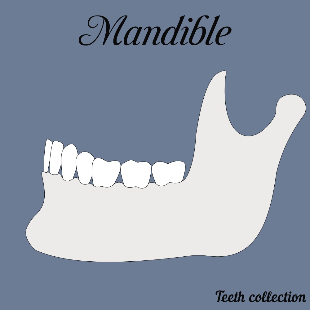
In a previous article, we reviewed the benefits of maxillofacial surgery utilizing augmented reality (AR) technology. However, as you may know, mandibular surgery is a major part of this surgical field, and consists of a wide variety of techniques and methods. Thankfully, AR is flexible, meaning that it can adapt to all kinds of situations and suit many different procedures. In today’s article, we will be talking about Mandibular Angle Split Osteotomy, which is basically a mandible reduction, but is differentiated from a mandible angle resection in the sense that it is more complete and, subsequently, extensive. We won’t get into too many of the technical and anatomical details with you all, but you can probably guess that it’s not an easy procedure, since it requires taking part of the mandibular bone out while being careful to maintain facial symmetry, and improving the overall aesthetic look of the patient’s face. For those of you who want a more technical description of the procedure, you can take a look at it HERE.

This technique has seen a lot of evolution since it was first instated, graduating from a purely manual procedure to one that utilizes robotic surgery to ensure precision and obtain the best results possible for the patient. Now, with the help of researchers from the Shanghai Jiao Tong University in Shanghai, China, a new add-on has been brought to the procedure. This system uses AR to decrease the invasiveness and overall risk of the surgery, and reduce the instance of long-lasting side effects from the surgery such as chronic pain from injury to the inferior alveolar nerve (a problem we have talked about AR’s possible role in HERE), or postoperative infection.

For the purpose of this study, surgeons without extensive mandibular surgery experience were brought in to test the system. The test consisted of providing 3D builds of the mandible constructed from CT scan data, with the intention of showing the surgeon how much bone should be extracted from the patient (in this case, animal models) and from where. The system was complete with a robotic “template” of sorts, which assessed the symmetry and precision of the cut before the surgeon made it. Four surgeries were performed on two “patients,” who were, in this case, dogs. This means that twenty tunnels were drilled into the mandible bones. The results showed that the average error margin at entrance and target points was 1.04 ± 0.19 and 1.22 ± 0.24 mm, which isn’t perfect, but is promising.

Angular error was also considered while assessing the results, and the difference between the planned and actual drilled tunnels was 6.69° ± 1.05°. The most important result, however, is that none of the dogs experienced any severe complications, suggesting that the system is a safe and practical method to implement in this type of surgery in the near future. Of course, this is something that must be approached with caution, but as with any other ‘futuristic’ method, results this good just mean we will be seeing this benefits on human models sooner rather than later, thanks to AR.


