
Epilepsy is a group of neurological disorders with one common denominator: the presence of epileptic seizures. These seizures are not produced by a temporary medical condition like low blood sugar, high fever, or hydroelectrolytic disturbance such as hyponatremia (low seric sodium). This group of pathologies have very diverse manifestations. Some people experience the classic symptoms of losing consciousness, collapsing, and shaking, but there can also be visual, auditory, olfactory, or behavioral changes during a seizure. Epilepsy can be debilitating for people, causing injuries when a person falls, and it’s not rare: at least 4.3 million adults were diagnosed with some form of epilepsy in 2013, according to the CDC. It’s usually manageable, but not always easy to diagnose or treat, and because of this, many people with epilepsy will be subjected to invasive and aggressive procedures.
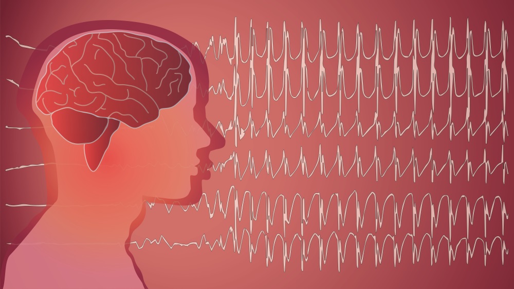
This is where Stereoelectroencephalography (SEEG) comes into play. SEEG is a procedure that consists of recording the electroencephalographic signals (just like a standard EEG) via depth electrodes implanted directly into the patient’s brain. It may sound radical and invasive, but it’s only used as a last resort for patients with severe epilepsy that is unresponsive to other treatments. It’s a last-ditch effort to avoid brain surgery,in many cases, but that does not mean it doesn’t come with risks. Because of this, researchers from the Department of Biomedical Engineering from the Tsinghua University’s School of Medicine in Beijing, China are proposing a new system that incorporates the use of augmented reality for the SEEG electrode implantation.
The system merges the use of video see-through AR (VAR) and spatial AR (SAR) with robotic surgery. This model in particular uses a projector camera system (PCS), which is attached to a robotic arm that serves as a surgical assistant to determine the specific insertion points into the operative field (the patient’s head, in this case). After that, the image is registered, and VAR is produced, merging the real-time video of the patient with the 3D preoperative planning model, which is built upon CT scan or MRI data. In addition to all this, SAR is implemented by projecting the electrode trajectories and local anatomical structures directly below the patient’s scalp to make a faster, more efficient, and safer system.

So, the big question here is obviously “does it work?” By measuring the error of registration related to the electrodes and the target points, all the measures were restricted into acceptable parameters, with no noticeable error. This was the objective of the feasibility study. Obviously, more studies are needed to determine the shortcomings of the system, but at least this provides a promising baseline study for the SEEG system for procedures that need accurate, deep placement of electrodes in even delicate locations in the body, all while projecting internal structural information to the surgeon and making their jobs easier and safer.

With this kind of system, surgeons can look at these procedures in an easy and intuitive way. We should expect more positive outcomes in the near future, thanks to augmented reality! What do you think? Let us know in the comments section below.


