
If you’re a frequent flyer here at ARinMED, it’s probably no surprise to you that neurosurgery is one specialty that uses augmented reality technology most often, and in some of the most creative ways. This makes sense–it’s one of the most complicated and dangerous specialties in terms of risk of harming the patient, and any tool that can help it be a little more accurate is welcomed. So far, we’ve talked about the usage of AR in preoperative planning, locating tumors, spinal surgery, and a whole lot more… but what about the actual brain surgery? Does AR have the potential to revolutionize that, too? Physicians from the Campus Virchow Klinikum in Berlin, Germany are ready to find out, and are harnessing the power of AR for intraventricular neuroendoscopy.

For the readers who are not doctors, intraventricular neuroendocsopy is a minimally-invasive (well, as minimally invasive as brain surgery can get, anyway) procedure in which the neurosurgeon accesses the brain through dime-sized drillings in the skull. The brain can then be viewed with the use of an endoscope attached to a high-resolution video camera. Sometimes, the endoscope is equipped with forceps and scissors. This procedure is most often used for tumor excision or biopsy. The brain is a delicate organ, and the struggle for neurosurgeons everywhere has always been the balance between completing the procedure while manipulating the actual brain tissue as little as possible in order to avoid injury and even death. The amazing thing about this procedure is that the recovery time is usually minimal.
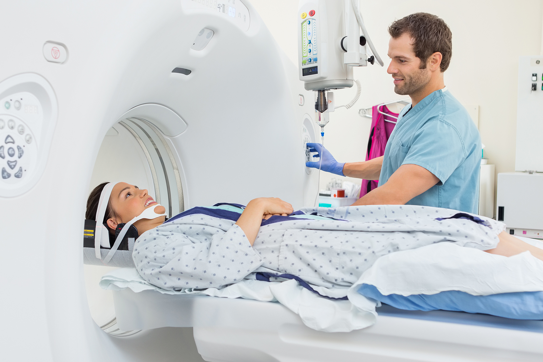
To test how AR technology could be applied to this technique, the researchers 3D printed a model of a head, which contained a lesion (tumor) in the right parietal lobe. This 3D model was viewed with a computer tomography (CT Scan), then segmented using the Scopis NovaPlan navigation software. Diagrams for the pre-planned region of interest and trajectory were superimposed over the endoscopic image using AR and a video-output of the endoscope. This superimposed image is basically a step by step guide to brain surgery. After testing the procedure on the 3D model head, 29 cases with cephalo spinal fluid (CSF) circulation pathologies were operated upon, including 18 cyst fenestrations, 10 biopsies, seven endoscopic third ventriculostomies, six stent replacements, and two shunt implantations. The results were amazing.
The accuracy of the system is astounding, with a deviation in the region of input midpoint of only 1.2 ± 0.4 mm, with diagnostic yield of 100%. The only complications observed in the patients were one postoperative hygroma and one insufficient fenestration, neither of which are uncommon in these sorts of procedures. The most important thing to take away here is that neuroendoscopy using AR is, at the moment, an accurate and feasible option. Of course, in order to be used in a clinical setting, it must undergo several levels of scrutiny to ensure its safety and efficacy, as well as trying to improve the precision.

While this experiment is great in theory, we’re not entirely sold yet. The results ultimately will depend on the surgeon’s skill level and experience with the AR technology, their confidence in the equipment, their comfort levels while using the tools, and many other factors. Is it really any better than the traditional intraventricular neuroendoscopy? If not, what needs to be improved? Scrutiny aside, we think that this is a very positive step for AR, and we hope to see it be perfected. What do you think about it? Let us know in the comments section below!
Source: https://link.springer.com/article/10.1007%2Fs00701-017-3152-x


