

It’s difficult to be a patient under suspicion of any type of cancer, and reaching the point where a biopsy needs to be performed, be processed, and then to await a result in order to know if you are or are not a patient with cancer. Several variables could have a positive or negative effect over this procedure, like the size of the sample, the location of the sample, and even the technique used to obtain this sample could all potentially affect the final outcome of this study. Take breast cancer, for example, where false negative results (the ones that worry us the most) are as common as 2.23%. A population of 988 patients underwent core needle breast biopsies, and 22 of these patients had a false negative result. This means that 22 of them were told that they didn’t have any cancer, but in fact, they did. Suddenly, 22 doesn’t sound like such a small number anymore, right?
With that in mind, it’s a common goal to develop more and more tools that enable us to decrease that number from 22 to zero. The goal is that each and every patient gets the right result at the right time, every time. That goal is shared by researchers from the Kebangsaan Malaysia University and the Putra Malaysia University, both located in Malaysia, and a system was proposed in order to create a 3D Augmented Reality (AR) Guidance Biopsy system, but in a different way than other studies (like this or this one), with researchers arguing that the already existent AR systems didn’t allow the physicians to perform a real-time localization of the tumor in order to improve the quality of the procedure.

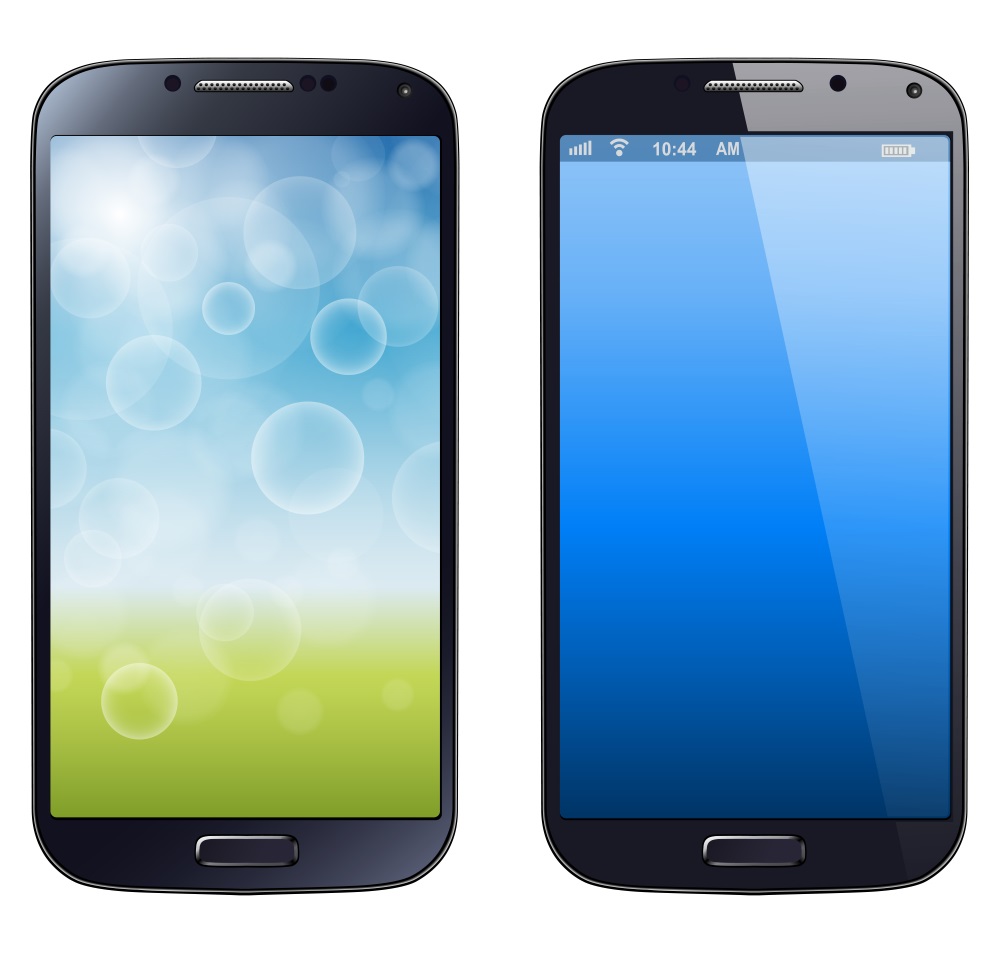
But how did they do it? This mobile AR visualization technique consists of different phases. First, the images are acquired through Computed Tomography (CT) or Magnetic Resonance Images (MRI), and after that, a 3D model is built using the DICOM format. This virtual 3D breast tumor model is applied to a breast phantom model and digital tracking through an open source vuforia tool, which allows interactive experiences with 3D objects and a final X-ray application in order to help locate the tumors beneath the skin of the model. Lastly, all of this information was uploaded into a smartphone device (specifically, a Samsung Galaxy S5) to perform the 3D tumor visualization. With this system, theoretically, the physician performing the biopsy will have a real-time, navigational system to perform the best possible biopsy! Plus, it’s a very inexpensive system!
This system was tested against stereotactic biopsy procedure with the task of targeting needle biopsies upon accurate tumor visualization in the training phantom. Even though the system has the potential to become a really great tool for this purpose, there were several limitations, such as the lack of a depth measuring, tracking method, and a very low frame rate limited the assessment of this tool. However, researchers have considered the implementation of ultrasound as a possible solution for all these issues, however, we can qualify this attempt as a great idea, since we know for a fact that it is indeed feasible with the right technology at a much more complicated field. We wonder what could be done about it now since ARCore is at the reach of everyone! It could probably improve the technical aspect of this procedure.
Certainly, this is a disappointment. We always hope (and are usually we are accustomed to) every single AR tool work as it was intended. However, that’s not always the case. But, we believe that this is an important idea – and we’d like to see what’s next in store for this research team in the near future!
What do you think? Do you have any suggestions to improve this study? Please let us know your thoughts in the comments section!


