You might think that “there’s an app for everything” is an exaggeration–but not for long! Doctors everywhere, especially in remote places with few resources, have always done the best they can with what they have, but this new development is taking that resourcefulness a step further. Brain surgery is a complex procedure that has traditionally taken lots of expensive planning, but neurosurgeons from the PLA General Hospital Hainan Branch and from the PLA General Hospital, both located in Hainan, China, are developing a novel system designed to minimize the cost of brain surgery for hospitals and patients alike.

Think you could plan a brain surgery with this?
One of the most important things to assess before performing brain surgery is, of course, the precise locations of intracranial lesions. The surgeon needs to know where to cut, after all. However, the MRI imaging information and other tests that are presently the gold-standard for making these assessments can be extremely expensive. The researchers wanted to make use of a cheaper tool, and what better place to start than something that so many people already have–a smartphone! There were almost 2.1 billion smartphone users in 2016, and that number is forecasted to jump to 2.87 billion by 2020. With this in mind, researchers thought that the iPhone (sorry, Android supporters!) could provide a low-cost navigation system to identify brain lesions and to construct the surface projections of the brain using augmented reality. The goal was to make these tests accurate and clinician-friendly.

Not all brain lesions are this obvious.
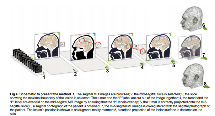
The study utilized 35 patients with brain lesions. Researchers used the augmented reality technology on the iPhone interface to view the lesion’s surface projection on the patient’s head. The first view was from the sagittal angle, taken and used in combination with the MRI picture of the patient using the FUSED app (Easy Tiger Apps LLC, App Store; Apple Inc.). With the patient’s augmented reality image in the background and the MRI image at the forefront, the images were manually adjusted to match each other by making the AR image transparent over the MRI image. Anatomical landmarks like the lips, nasal tip, curve of the head, and external occipital protuberance were used to center and adjust the images, resulting in an AR model that could be used to pinpoint the location of the brain tumor.
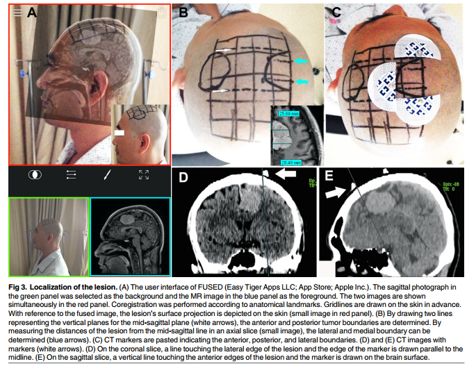
While you’ll have to consult the article for specific technical data (and we welcome you to do so!), the gist of it is that researchers found that 97.7% of the markers demonstrated a high level of accuracy, with a median margin of error of 2.90 mm. The maximum error was 4.2 mm, which is certainly not negligible, especially in neurosurgery, but the preliminary results are still incredibly promising, even if there’s still work to be done.
Here in ARinMED, we believe that all great advancements start with a creative mind!
The simplicity here is what’s so amazing. There’s no special equipment and no specially designed app–just a team of talented, bright people using smartphones in a creative new way. We think that’s pretty incredible, and the implications are amazing–the more high-quality medical work that can be done on a low budget, the higher the standard of care will be for regions who can’t afford more expensive machinery. What do you think? Let us know in the comments section!
Source: https://www.ncbi.nlm.nih.gov/pmc/articles/PMC4959690/pdf/pone.0159185.pdf
Categories: Neurosurgery, Surgery, Radiology, Practice based apps.


