If you have been reading our articles lately, you may have noticed that we are focusing more and more into ‘real time’ mechanisms to deliver Augmented Reality into several medical fields. This is especially true when talking about medical emergencies, because even though we believe that Augmented Reality is truly helpful for elective procedures, there are several methods that enable planning surgeries (with AR becoming one of those methods) and performing them successfully. The real strength of it – the place where it will deliver a big difference is right in the emergency room – when there is no time for planning, only immediate actions, and there’s no place with more action than the emergency trauma room.
Some of the most common fractures are the ileocecal joint fracture and superior pubic ramus fracture. To repair them, patients undergo a surgery that is considered high-risk due to the fixation that needs to be done. The surgeon passes a metal wire (Kirschner wire) through muscles and bone fragments by using pre-operative X-ray imaging, but the 2D nature of these images sometimes makes it difficult to portray every structure, therefore making complications such as injury or even life-threatening risks to patients. Researchers from the Computer Aided Medical Procedures Department and the Trauma Department at both the Johns Hopkins University and the Universität München in Munich, Germany have proposed a new Augmented Reality system that could be viewed as a solution for this matter.

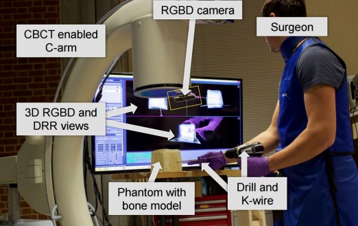
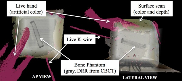
A 3D Augmented Reality System that uses a CBCT (Cone Beam Computed Tomography) enabled C-arm, which is a low radiation device, in combination with a color and depth (RGBD) camera. By merging both elements, a 3D reconstruction and visualization of the operative field in real time is obtained, and that model is portrayed in Augmented Reality to give the surgeon both physical and anatomical information from any point of view, in thus, allowing the surgeon to avoid the previously mentioned complications. Registration and calibration is required to focus the system on the relevant structures for the surgery and to discard the noisy background, but it’s a practical and easy process that can be done prior to the surgery and doesn’t limit the real-time properties of the system (as seen below).
The system was tested using a bone model phantom, with 7 trained surgeons performing the surgical procedure at superior pubic ramus bone phantoms. Each surgeon performed the same surgery using the standard equipment and the Augmented Reality system in random order, where the procedure lasted 9.9 min and 4.1 min respectively, which is a statistically significant improvement (p <0.05). Other compared elements included the number of acquired X Ray images (40.9 to 2.1), X Ray dose (4.43 to 1.60 cGycm2), and surgical task load (43.5 to 17.6), which are all statistically significant improvements over the standard procedure (p < 0.05).
We believe that the results of the study are very clear, the strong build of this systems enables it not only to be ‘not inferior’ to the standard procedure, but to overcome it in many ways, making it a faster and safer equipment compared to what we are currently using in our hospitals every day. Of course, more studies are needed in order to analyze the prospect of standardizing this technology, but Augmented Reality is surely proving its value.
Do you think there are other applications for this kind of technology? Please share with us in the comments section!


