
Coronary computed tomography angiography (CTA) is an imaging test designed to facilitate a nonsurgical procedure called a percutaneous coronary intervention (PCI). PCI, also called a coronary angioplasty, is used to treat the narrowing of coronary arteries in patients with coronary diseases. It involves inflating a soft catheter with a balloon on its end inside the heart, and a tube called a stent into the heart valve to restore normal blood flow. This procedure is designed to save the lives of people who have had or are at a high risk for heart attacks, but it can be invasive.
A CTA can predict not only the likelihood of successful PCI, but also the clinical benefit of the revascularization, which allows physicians to decide whether the procedure would improve a patient’s quality of life enough to warrant the procedure. While the procedure is not a surgical one, there are always risks involved, and physicians need to weigh those risks when deciding whether to subject a patient to medical intervention. The CTA imaging allows doctors to more quickly and accurately make those decisions.
The CTA can allow physicians to characterize coronary atherosclerotic plaques and to determine the extent of artery calcification, vessel tortuosity (when a vessel is twisted), and the presence of multiple clots or blockages. It can also allow physicians to view a 3-dimensional image of coronary vessel trajectories, allowing them to more accurately and safely insert stents and other medical instruments into the narrow walls of the arteries. Sadly, however, this imaging technique is still limited by economic and technical factors.
Researchers from the Department of Interventional Cardiology and Angiology of the Institute of Cardiology at Warsaw, Poland have developed a unique way to provide the coronary CTA with the flexibility of an AR wearable device (in this case, Google Glass). The patient that this method was first used upon was a 49-year old hypertensive patient. This person had already had a coronary artery bypass grafting and PCI, but was still experiencing grade III angina (in layman’s terms, the patient had chest pains even with only minimal exertion, such as walking through a store or up a flight of stairs.) The cardiac MRI showed a preserved left ventricular ejection fraction, otherwise known as diastolic heart failure. Diastolic heart failure simply means that the problem occurs when the heart should be filling with blood, as opposed to pumping it out. The patient also presented with a reversible perfusion defect in the right coronary artery (RCA) territory, which means that under stress, certain areas of the heart experienced reduced blood flow. In short, the patient was in pain, and areas of the heart were not filling properly with blood.
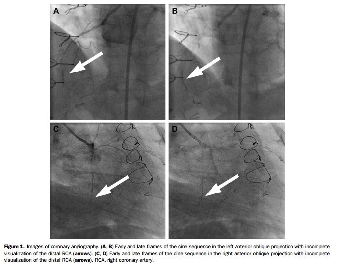
After the PCI was performed, coronary angiography showed the presence of a stent in the left circumflex artery (LCA), the presence of a left internal mammary artery graft (a procedure that allows the heart to bypass the areas that don’t work and supplement their function with areas of the heart that do work) in the obstructed artery, and chronic blockages resulting in incomplete filling of the distal (farthest away from the center of the body) heart vessel (figure 1 on the article). A coronary CTA was performed, which revealed severe calcification near the blockages, more severe than the doctors had previously thought. Once more, to simplify, the patient had blockages in the heart which needed to be addressed before it was too late.
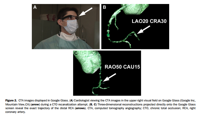
3D CTA reconstructions of the damaged areas of the patient’s heart were selected using the syngo.via software (SIEMENS) and projected onto the Google Glass for display. This allowed the operators to view the projections of the patient’s heart and scans with Google Glass, and to verify the necessary trajectory of the guide wire. The PCI was successful: two drug-eluting stents were implanted without any reported complications.
This case, among many others, demonstrates the potential of AR wearable dispositives in the medical field. Used in the catheterization laboratory, it can allow for better planning and guidance of interventional procedures, making them less risky and more effective. Access to AR technology is getting easier as the technology develops, and we would love to see it reach more hospitals across the world. We at ARinMED believe that AR/MR technology have a great potential to optimize countless procedures in the medical field. Tell us what you think in the comments section!
SOURCE: http://www.cpsmd.com.cn/data/upload/ueditor/20160715/5788c1b55c0b2.pdf


