‘Primum non nocere’, or ‘first, do no harm’, is one of the phrases that is dogmatic in medicine – we are able to do almost every conceivable procedure, but first of all we must guarantee that we are not doing more harm than good. This applies to almost every aspect of medicine. This phrase usually comes to mind in complex situations, and it’s so important that it even spawned a new medical field (bioethics). It is one of those things that is in every (righteous) physician’s head, almost all the time when practicing medicine, consciously or unconsciously.

When talking about radiology, a physician does not order unnecessary studies because it is known that every single X-Ray or CT Scan comes with a hefty dose of radiation and even in the long run, it is harmful. Studies have been revised where complex 3D models are built from these kinds of studies but, if there’s a way to cut the quantity of radiation, that would be ideal. We wouldn’t want to have great 3D models at the expense of radiating patients 2, 3, 4, 5 times more than what is required, right? Let’s remember that just one CT Scan packs the same amount of radiation than 200 X-Rays, and sadly most doctors underestimate those dangers. Fortunately, technology can do something about it.

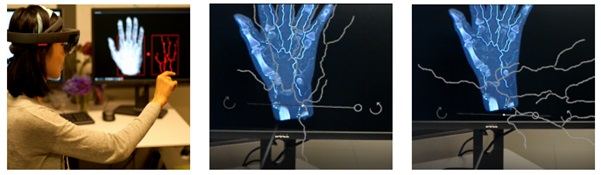
One very recent study, which was made in collaboration with researchers from the University of Washington in Seattle, US along with researchers from the Kyushu University, the National Institute of Informatics, and Keio University in Japan. The proposed system skips the use of CT Scan, and instead embraces the use of Stereo X-Ray to reconstruct and visualize 3D blood vessels (even in the presence of fat or muscle tissue) in order to portray them in Augmented Reality through the Microsoft HoloLens.
Stereo X-ray is not a new procedure. It started in the mid 90’s, and it consists on obtaining the imaging by using various X-ray sources. For the purpose of this study, the imaging was obtained with the same X-ray tube from different positions. Together, with the use of endovenous contrast allowed to obtain the vascular images through this method. These vascular images were segmented and matched between both X-ray images, and then using the differences between both of them a 3D model was easily rendered that was exported directly to the Microsoft HoloLens platform, where users can interact with it openly, rotate, and explore it in every way needed.
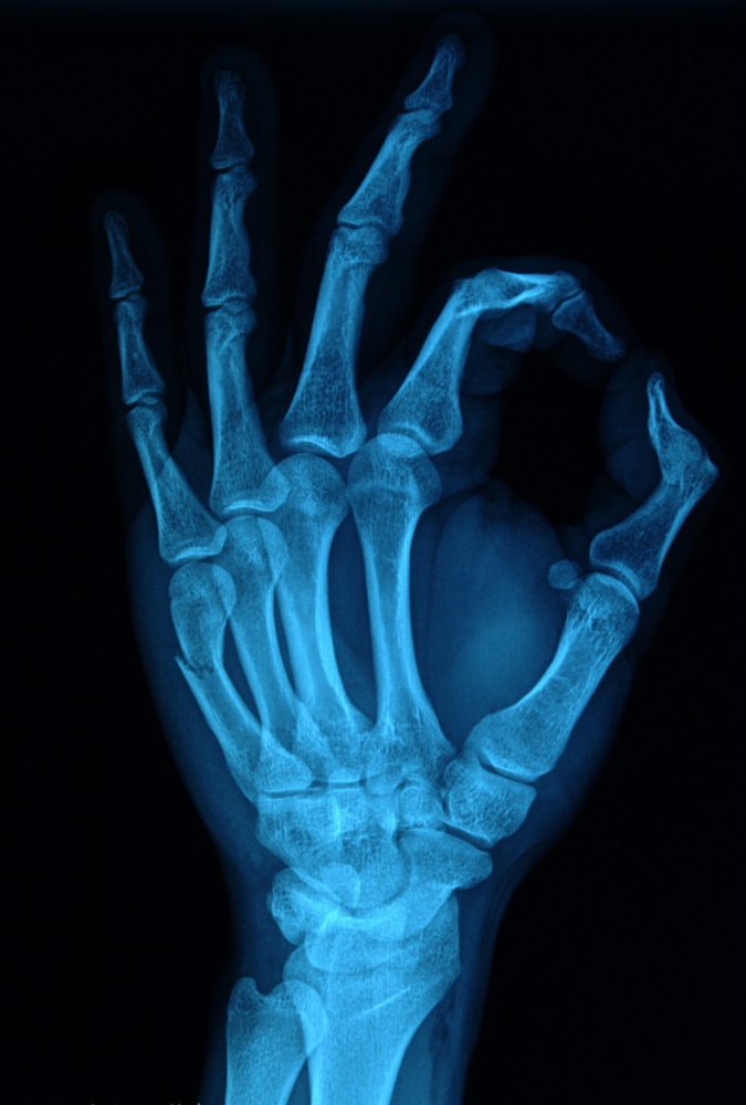
For this experiment, the system was used in a human hand, which is an especially difficult structure because of the contrast differences with the bone, but ultimately the system accomplished its objective very well, and the visualization of blood vessels was possible as you can see in the image below.
This is an important step forward to developing AR systems that are less dependent to heavy-radiation doses, we also know from previous experience with Stereo X-ray that almost any kind of vascular imaging can be done. We picture this system working very well for vascular procedures that need pre-operative planning, such as Aneurysms or arteriovenous malformations, elevating the sensitivity and sensibility of this radiological technique without compromising the health of the patient, keeping radiation levels low. All of this is thanks to the creativity of these researchers and the power of Augmented Reality.
What do you think about Stereo X-ray? Do you think there are other uses for this technique in the AR setting? Please share them with us in the comments section!
Source: https://arxiv.org/abs/1709.07551


